Echo-Guided Biopsy⁚ A Comprehensive Guide
This guide explores echocardiographic guidance in biopsies, a technique using ultrasound to precisely target tissue samples. It details the procedure, advantages over other methods, potential risks, and interpretation of results. We will examine its application in various biopsy types and future advancements in this crucial medical field.
Echo-guided biopsy, a minimally invasive procedure, leverages real-time echocardiographic imaging to precisely guide a needle to the target tissue for sampling. This sophisticated technique offers significant advantages over traditional biopsy methods, particularly in accessing challenging anatomical locations. The procedure’s precision minimizes the risk of damage to surrounding healthy tissues and organs. Echocardiography’s real-time visualization allows for immediate adjustments during the biopsy, enhancing accuracy and safety. This approach is particularly valuable when targeting lesions within the heart or nearby structures, where traditional imaging methods may prove insufficient. The integration of ultrasound technology with biopsy procedures significantly improves the diagnostic process, leading to more accurate diagnoses and effective treatment planning. The advantages of using echo guidance include real-time imaging capabilities, portability, and widespread availability, which contribute to its effectiveness as an imaging modality in various medical settings. The combination of real-time imaging and precise needle guidance makes echo-guided biopsy a superior choice for many biopsy procedures.
Types of Image-Guided Biopsies⁚ Ultrasound vs. CT vs. Echo
Image-guided biopsies employ various imaging modalities to precisely locate and sample tissue. Ultrasound, a widely used technique, uses sound waves to create real-time images, guiding needle placement for biopsies of superficial lesions. Computed tomography (CT), utilizing X-rays, provides detailed cross-sectional images, ideal for biopsies of deeper structures or those requiring precise targeting within complex anatomy. Echo-guided biopsy, specifically using echocardiography, excels in visualizing cardiac structures and surrounding tissues. This makes it uniquely suited for biopsies of the heart, pericardium, or adjacent areas. The choice of imaging modality depends on the target location, size, and accessibility of the lesion. While ultrasound is often preferred for superficial lesions due to its ease of use and real-time imaging, CT is valuable for deeper lesions where more precise localization is required. Echocardiography offers superior real-time visualization for cardiac and related structures, making it the preferred method for these specific applications. Each technique presents unique advantages, necessitating careful consideration of the individual patient’s needs to select the most appropriate approach. The selection process takes into consideration the location, size, and accessibility of the targeted lesion.
Ultrasound-Guided Biopsy Techniques and Applications
Ultrasound-guided biopsy leverages real-time imaging to precisely guide needle placement, minimizing invasiveness and maximizing accuracy. Techniques vary depending on the target tissue and biopsy type. For superficial lesions, a fine-needle aspiration biopsy (FNAB) may suffice, extracting cells for cytological examination. For deeper or larger lesions, a core needle biopsy may be necessary, obtaining tissue cylinders for histological analysis. Applications are extensive, encompassing various organs and systems. Breast biopsies, for instance, frequently employ ultrasound guidance to target suspicious masses or microcalcifications. Thyroid and liver biopsies also benefit from ultrasound’s ability to visualize and navigate through these complex anatomical regions. Ultrasound-guided biopsies are routinely performed for musculoskeletal lesions, guiding needle placement within joints, muscles, and bones. In interventional radiology, ultrasound assists in procedures such as draining fluid collections (cysts or abscesses) under image guidance, minimizing risks. The versatility of ultrasound-guided biopsy makes it a cornerstone technique in numerous medical specialties, offering a minimally invasive approach to tissue sampling for diagnosis and treatment planning. Its real-time imaging capability allows for continuous monitoring during the procedure, ensuring accurate needle placement and minimizing complications.
Echocardiographic Guidance in Percutaneous Procedures
Echocardiography plays a pivotal role in guiding percutaneous procedures, offering real-time visualization of cardiac structures and surrounding tissues. Its application extends beyond simple imaging; it actively directs instruments during interventions. In percutaneous biopsy of cardiac masses or lesions, echocardiography provides crucial anatomical information, ensuring accurate needle placement while minimizing the risk of complications such as perforation or bleeding. Real-time imaging allows for dynamic adjustments during the procedure, adapting to variations in cardiac anatomy and ensuring precise targeting of the lesion. The technique is particularly valuable in accessing difficult-to-reach areas within the heart, such as septal lesions or those located in the right ventricle. Furthermore, echocardiographic guidance facilitates the safe and efficient placement of catheters for various therapeutic interventions, including the drainage of pericardial effusions or the deployment of pacemakers. By providing a clear image of the heart’s internal structures and the surrounding vasculature, echocardiography enables precise and controlled manipulation of instruments, enhancing the safety and efficacy of these procedures. The use of echocardiography in percutaneous procedures improves the accuracy and safety of various cardiac interventions, making it an indispensable tool in modern cardiology.
Advantages of Echo-Guided Biopsy
Echo-guided biopsy offers several key advantages over other biopsy techniques. Its real-time imaging capability allows for precise needle placement, maximizing the likelihood of obtaining a representative tissue sample while minimizing the risk of injury to surrounding structures. This is particularly crucial in areas with complex anatomy or where the target lesion is small or difficult to access. The procedure is minimally invasive, often requiring only local anesthesia, leading to shorter recovery times and reduced patient discomfort compared to open surgical biopsies. The portability of ultrasound equipment allows for biopsies to be performed at the patient’s bedside or in various clinical settings, eliminating the need for transfer to a specialized imaging suite. The real-time feedback provided by ultrasound allows for immediate adjustments during the procedure, ensuring optimal needle placement and sample acquisition. This real-time guidance also reduces the number of needle passes required, further minimizing the risk of complications. Moreover, the cost-effectiveness of echo-guided biopsy, combined with its high accuracy and reduced invasiveness, makes it an attractive option for many patients and healthcare providers. The procedure’s speed and efficiency contribute to optimized workflow in busy clinical environments.
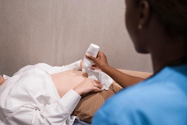
Procedure Details⁚ Preparation, Process, and Recovery
Preparation for an echo-guided biopsy typically involves a brief consultation with the physician to discuss the procedure, potential risks, and answer any questions. A medical history is reviewed, and any relevant allergies or medications are noted. Depending on the biopsy site and physician preference, local anesthesia may be administered to numb the area. The process itself involves the placement of a transducer on the skin, allowing the physician to visualize the target area in real-time using ultrasound. A thin needle is then carefully guided to the suspicious area under ultrasound visualization. Once the needle is in place, tissue samples are collected. The entire procedure usually takes only a few minutes. Following the biopsy, a small bandage is applied to the puncture site. Patients may experience mild discomfort or bruising, which usually resolves within a few days. Specific post-procedure instructions, such as avoiding strenuous activity or monitoring for any signs of infection, will be provided by the physician. Recovery time is generally short, allowing patients to resume their normal activities within a day or two. The collected tissue sample is then sent to a pathology lab for analysis.
Risks and Complications Associated with Echo-Guided Biopsy
While generally safe, echo-guided biopsy carries potential risks and complications, although these are infrequent. Minor bleeding or bruising at the puncture site is common and usually resolves quickly. Infection is a possibility, though minimized by sterile techniques. Rarely, a hematoma (blood clot) may form, requiring further medical attention. Pneumothorax (collapsed lung) is a risk, particularly with biopsies near the lungs, necessitating close monitoring. The needle might accidentally puncture an adjacent organ or blood vessel, leading to complications depending on the location. These occurrences are uncommon with skilled practitioners and appropriate imaging guidance. The accuracy of the biopsy depends on several factors including the size and location of the target lesion and the expertise of the operator. In some instances, an insufficient tissue sample may be obtained, necessitating a repeat procedure. The patient’s overall health and any pre-existing conditions can also influence the risk profile. Pre-procedure discussions with the physician are crucial to understanding and addressing these potential risks, ensuring informed consent and managing expectations.
Interpreting Results⁚ What to Expect After the Biopsy
Following an echo-guided biopsy, the retrieved tissue sample is sent to a pathology laboratory for analysis. The pathologist examines the tissue under a microscope to determine its cellular structure and identify any abnormalities. The results typically take several days to a week, depending on the complexity of the analysis and the laboratory’s workload. The reporting physician will communicate the findings to the patient and their referring physician, explaining the significance of the results in the context of their overall health. A positive result indicates the presence of abnormal cells, potentially cancerous or indicative of other diseases. This may necessitate further investigations and treatment. A negative result indicates no evidence of disease in the examined tissue sample. However, it’s crucial to remember that a negative result doesn’t always rule out the possibility of disease elsewhere in the body. The interpretation of biopsy results requires medical expertise, and patients should discuss their results with their healthcare provider to understand their implications and receive appropriate guidance on follow-up care. Clear communication and careful explanation of the results are paramount to patient understanding and informed decision-making.
Comparison of Echo-Guided Biopsy with Other Methods
Echo-guided biopsy offers several advantages compared to other image-guided techniques. Unlike CT-guided biopsies, which involve ionizing radiation, echo-guided procedures are radiation-free, minimizing patient exposure to harmful effects. Compared to ultrasound-guided biopsies, echocardiography provides real-time visualization of the heart and surrounding structures, crucial for procedures involving cardiac tissue. While both ultrasound and echo use sound waves, echocardiography’s higher resolution and ability to visualize moving structures enhances precision, particularly in challenging anatomical locations. The portability of echocardiography equipment also allows for bedside procedures, reducing the need for patient transport. However, echo-guided biopsies might be limited by operator expertise and the quality of the echocardiographic images, which can be affected by factors such as patient body habitus and underlying medical conditions. The choice of method depends on the specific clinical scenario, the target tissue, and the available resources. The advantages of echo guidance in terms of real-time imaging and precision, coupled with its radiation-free nature, make it a valuable tool in specific situations, but its application should be considered in relation to other established methods.
Role of Echo Guidance in Specific Biopsy Types (e.g., Breast, Thyroid)
While not as prevalent as ultrasound or CT guidance for breast or thyroid biopsies, echocardiography plays a specialized role in specific situations. In breast biopsies, echo guidance might be considered for lesions near the chest wall or those with complex anatomical relationships to surrounding structures, offering a clearer view than standard ultrasound alone. Similarly, in thyroid biopsies, echo guidance can improve precision when dealing with deep-seated nodules or those adjacent to critical vascular structures. The real-time imaging capabilities of echocardiography allow for precise needle placement, minimizing the risk of complications such as bleeding or pneumothorax. However, the use of echo-guidance in these contexts remains less common than other modalities due to the specialized expertise required and the availability of equipment. For routine breast and thyroid biopsies, ultrasound-guided procedures remain the standard of care. Echo-guided biopsy in these areas is a niche application, suited for complex cases or when the advantages of real-time cardiac imaging outweigh the specialized expertise and equipment requirements.
Future Directions and Advancements in Echo-Guided Biopsy
The field of echo-guided biopsy is poised for significant advancements. Integration of advanced imaging techniques, such as 3D and 4D echocardiography, promises enhanced visualization and precision. This could lead to smaller needle sizes, reducing patient discomfort and the risk of complications. Furthermore, the development of sophisticated software for image fusion and navigation systems will improve the accuracy of needle placement and minimize the need for multiple passes. Artificial intelligence (AI) is also emerging as a powerful tool. AI-powered algorithms can analyze images in real-time, providing clinicians with valuable insights and assisting in the identification of target lesions. This will likely result in faster procedure times and improved diagnostic accuracy. Miniaturization of transducers and improved sensor technology will enhance the portability and ease of use of echo-guided biopsy systems, potentially enabling procedures in more diverse settings. The integration of robotic assistance could further improve precision and reduce operator dependence. These technological advancements hold the promise of making echo-guided biopsy safer, more efficient, and more widely accessible.